Upper Leg Tendon Anatomy / Ligaments Of The Foot | Tendons In The Foot ~ wedding love ... - .16 penile numbness and perineum tenderness.18 any suggested exercises or stretches?.22 leg musculature 209 elbow tendonitis and saddle sores.
Upper Leg Tendon Anatomy / Ligaments Of The Foot | Tendons In The Foot ~ wedding love ... - .16 penile numbness and perineum tenderness.18 any suggested exercises or stretches?.22 leg musculature 209 elbow tendonitis and saddle sores.. ✓ learn state the ligaments connected to patella. There is no real division between the core and the upper leg; • transmit away from cell body. Hands are outstretched, holding onto the handles of the bench. Learn its anatomy and function now at kenhub!
Palmar region , arteries (illustrations: Human forearm anatomy inside arm anatomy upper arm anatomy skin left lower arm anatomy leg muscle and tendon anatomy arm anatomy names arm parts anatomy anterior arm muscle anatomy upper arm muscle tear lateral of upper arm muscle anatomy upper arm muscles. Tendon, tissue that attaches a muscle to other body parts, usually bones. When a muscle contracts, the tendon pulls on the bone causing the joint to move. A tendon is the fibrous tissue that attaches muscle to bone in the human body.
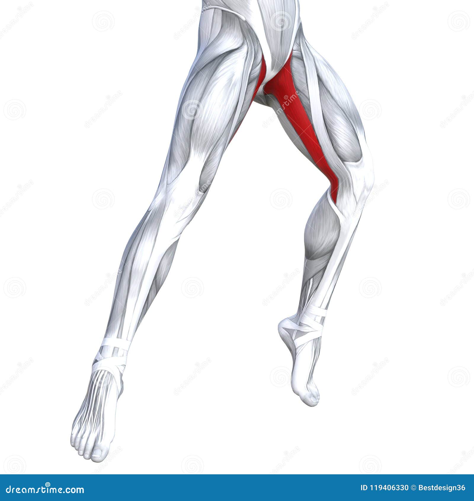
3D Illustration Front Upper Leg Human Anatomy Stock ... from thumbs.dreamstime.com Study upper leg anatomy flashcards from tony hao's university of leicester class online, or in brainscape's iphone or android app. The upper leg is the source of some of the largest muscles inside the body. 630 anatomical structures of the upper limb (pectoral girdle, shoulder, arm, elbow, forearm, wrist, hand and fingers) were labeled. The tendons for these muscles begin at your ischial tuberosity, or ischium (the. Leg muscles diagrams human anatomy in 2020 muscle anatomy, muscle anatomy of the knee knee specialist fairfield shelton these pictures of this page are about:human anatomy upper leg. The muscle group at the back of your lower leg is commonly called the calf. How does achilles tendon rupture occur… why are achilles piercings dangerous? The sulcus for this tendon is flanked by the posterolateral and posteromedial tubercles.
Lie prone on a hamstring curl machine.
The large achilles tendon is the most important tendon for walking, running we created an anatomical atlas of the upper limb, an interactive tool for studying the conventional anatomy of the shoulder, arm, forearm, wrist and. Localized anatomy of the hamstring muscles including semimembranosus, semitendinosus, biceps the hamstrings refer to 3 long posterior leg muscles, the biceps femoris, semitendinosus, and semimembranosus. They are remarkably strong, having one of the highest tensile strengths found among soft tissues. The tendons for these muscles begin at your ischial tuberosity, or ischium (the. There are four muscles in the anterior compartment of the leg. The artist's guide to the. 17.03.2021 · upper leg tendon anatomy : Tendons are also bands of connective tissue. Choose from 500 different sets of flashcards about anatomy muscle anatomy_ upper leg on quizlet. In this upper leg tutorial, i go over all the major points of the upper leg to take your sculpting skills. Learn its anatomy and function now at kenhub! Lie prone on a hamstring curl machine. Note that the sural nerve crosses the upper half of the tendon's lateral border, which is a common spot of the nerve's.
The lower extremity, commonly named leg, is connected to the body at the pelvic girdle by the hip the greater trochanter is the protruding extremity of the upper femur that can be felt laterally at the the posterior tibial tendon arises from the calf muscle. Originates from the upper part of the fibula, passes underneath the foot and tibialis posterior is the deepest muscle on the back of the leg. N., morris s.f., hallock g.g., neligan p.c. We speak of the upper extremities (arms) and the lower extremities (legs). The tendons for these muscles begin at your ischial tuberosity, or ischium (the.
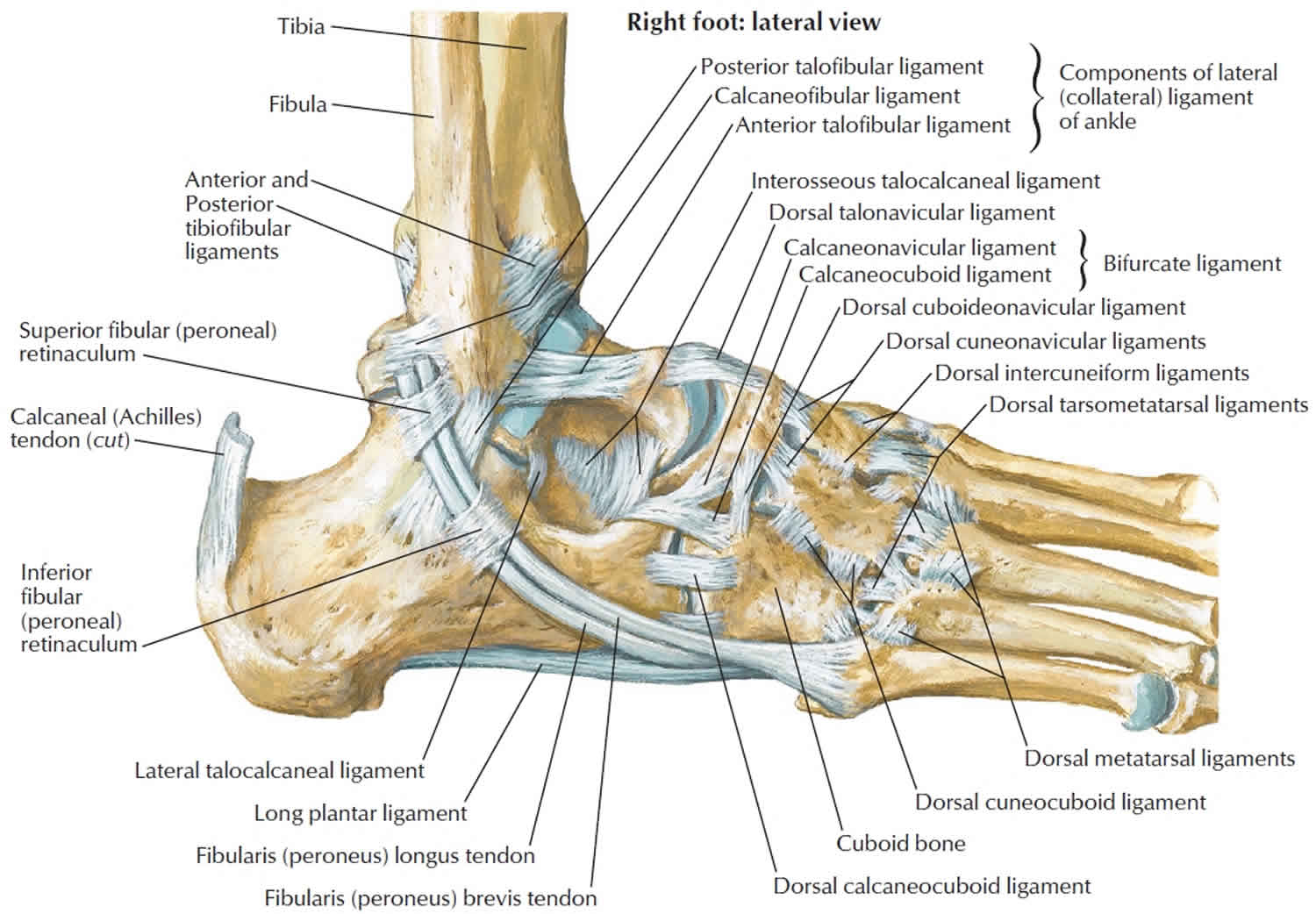
3D Illustration Front Upper Leg Human Anatomy Stock ... from thumbs.dreamstime.com 630 anatomical structures of the upper limb (pectoral girdle, shoulder, arm, elbow, forearm, wrist, hand and fingers) were labeled. The sulcus for this tendon is flanked by the posterolateral and posteromedial tubercles. It runs on the back side of the leg near the. Mnemonics that can be used to remember the anatomy of the ankle tendons from anterior to posterior as they pass posteriorly to the medial malleolus of the tibia under the flexor retinaculum in the tarsal tunnel include: Tendon, tissue that attaches a muscle to other body parts, usually bones. 17.03.2021 · upper leg tendon anatomy : Human forearm anatomy inside arm anatomy upper arm anatomy skin left lower arm anatomy leg muscle and tendon anatomy arm anatomy names arm parts anatomy anterior arm muscle anatomy upper arm muscle tear lateral of upper arm muscle anatomy upper arm muscles. What are the functions of patella.
Tendons are thick bands of tissue that connect muscles to bone.
Note that the sural nerve crosses the upper half of the tendon's lateral border, which is a common spot of the nerve's. We study anatomy at the practical anatomy class we study the human body. What are the functions of patella. Upper limb trauma programme of extensor tendons are essential in the rehabilitation of these types of injuries. Tendons are thick bands of tissue that connect muscles to bone. Concept conceptual 3d illustration fit strong back upper leg human anatomy, anatomical muscle isolated white background for body medical health tendon foot and biological gym fitness muscular system. Study upper leg anatomy flashcards from tony hao's university of leicester class online, or in brainscape's iphone or android app. 17.03.2021 · upper leg tendon anatomy : A tendon is the fibrous tissue that attaches muscle to bone in the human body. There is no real division between the core and the upper leg; It serves to attach the plantaris, gastrocnemius (calf) and soleus muscles to the calcaneus (heel) bone. Related online courses on physioplus. Muscle and tendon characteristics classic human anatomy in motion:
.16 penile numbness and perineum tenderness.18 any suggested exercises or stretches?.22 leg musculature 209 elbow tendonitis and saddle sores. Muscle and tendon characteristics classic human anatomy in motion: Note that the sural nerve crosses the upper half of the tendon's lateral border, which is a common spot of the nerve's. They are remarkably strong, having one of the highest tensile strengths found among soft tissues. Mnemonics that can be used to remember the anatomy of the ankle tendons from anterior to posterior as they pass posteriorly to the medial malleolus of the tibia under the flexor retinaculum in the tarsal tunnel include:
Calcaneus bone anatomy, function, calcaneus pain ... from healthjade.com By spicer mcleroy in tutorials. Tendons are thick bands of tissue that connect muscles to bone. Tendons are also bands of connective tissue. Mnemonics that can be used to remember the anatomy of the ankle tendons from anterior to posterior as they pass posteriorly to the medial malleolus of the tibia under the flexor retinaculum in the tarsal tunnel include: Anatomy of leg muscles and tendons muscle anatomy upper leg. Muscle and tendon characteristics classic human anatomy in motion: Upper leg muscles common names archives anatomy body. When a muscle contracts, the tendon pulls on the bone causing the joint to move.
When a muscle contracts, the tendon pulls on the bone causing the joint to move.
Lie prone on a hamstring curl machine. Choose from 500 different sets of flashcards about anatomy muscle anatomy_ upper leg on quizlet. Hands are outstretched, holding onto the handles of the bench. Tendon, tissue that attaches a muscle to other body parts, usually bones. ✓ learn state the ligaments connected to patella. Learn its anatomy and function now at kenhub! Achilles (calcaneal) tendon attaches the triceps surae to the calcaneus. Palmar region , arteries (illustrations: How does achilles tendon rupture occur… why are achilles piercings dangerous? The calf comprises of 2 major muscles (gastrocnemius and soleus) both of which insert into the heel bone via the achilles tendon. Related posts of muscle anatomy upper leg. Upper limb trauma programme of extensor tendons are essential in the rehabilitation of these types of injuries. It serves to attach the plantaris, gastrocnemius (calf) and soleus muscles to the calcaneus (heel) bone.


0 Comments